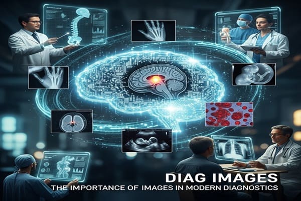
In today’s fast-paced medical world, visuals aren’t just helpful—they’re essential. Diag images, from X-rays to MRIs, have become the backbone of modern diagnostics. They give doctors a window into the human body, revealing insights that were once almost impossible to detect.
These images do more than show structures—they help healthcare professionals connect symptoms to accurate diagnoses, plan effective treatments, and monitor patients’ progress. Whether you’re a medical professional or a curious patient, understanding diag images is becoming increasingly important. Let’s take a closer look at this fascinating intersection of technology and medicine.
The Evolution of Medical Imaging
Medical imaging has come a long way. In the early days, doctors relied on basic observations and physical exams, supplemented occasionally by simple X-rays. These were groundbreaking at the time but offered limited insight into internal structures.
Key milestones in medical imaging:
-
X-rays: Introduced a way to see bones and certain tissues without surgery.
-
CT scans: Provided detailed cross-sectional images of organs, improving diagnostic accuracy.
-
MRI scans: Offered high-resolution views of soft tissues like the brain and spinal cord without radiation exposure.
-
Digital imaging: Made images clearer and easier to share, while speeding up diagnosis.
-
AI integration (today): Promises faster, more precise interpretations and the detection of subtle abnormalities that might be missed by the human eye.
This journey from basic observation to AI-assisted diagnostics has fundamentally changed patient care, making it faster, safer, and more precise.
Why Diag Images Matter
Diag images are more than just pictures—they’re powerful tools that impact nearly every aspect of healthcare. Here’s why they matter:
-
Enhanced accuracy: High-quality images help doctors detect conditions early, improving treatment outcomes.
-
Better treatment planning: Visualizing internal structures allows for personalized approaches to care.
-
Non-invasive options: Many imaging methods reduce the need for invasive procedures, making the patient experience safer and more comfortable.
-
Monitoring progress: Doctors can track disease progression and adjust treatments in real-time.
With these advantages, diag images have moved from being optional tools to indispensable instruments in modern medicine.
Common Types of Diag Images
Different situations call for different imaging techniques. Here’s a breakdown of the most common diag images:
-
X-rays: Ideal for examining bones, detecting fractures, and spotting certain tumors.
-
CT (Computed Tomography) scans: Offer detailed cross-sectional images of organs and tissues, giving a more complete picture than X-rays.
-
MRI (Magnetic Resonance Imaging): Provides high-resolution images of soft tissues like the brain, muscles, and joints without exposing patients to radiation.
-
Ultrasound: Uses sound waves to generate real-time images, commonly used in pregnancy and organ assessment.
-
PET scans: Detect metabolic activity in the body, helping in oncology and neurology.
Each imaging type has its strengths and is chosen based on the medical question at hand.
Applications Across Medical Specialties
Diag images are not limited to one area—they touch nearly every branch of medicine:
-
Oncology: Imaging detects tumors, measures size, and tracks treatment response.
-
Cardiology: Echocardiograms and CT scans reveal heart structure, blood flow, and valve issues.
-
Orthopedics: MRIs help identify joint injuries, soft tissue damage, and degenerative conditions.
-
Neurology: CT and MRI scans are crucial for diagnosing strokes, brain injuries, and neurological disorders.
-
Pediatrics: Imaging monitors growth and development safely, adjusting techniques to suit younger patients.
These applications show just how integral diag images are to improving both diagnosis and treatment outcomes.
Challenges and Limitations
Despite their benefits, diag images are not without hurdles:
-
Accessibility: Not all healthcare facilities can afford advanced imaging technology.
-
Interpretation complexity: Even experienced radiologists face challenges in analyzing images due to anatomy variations or overlapping structures.
-
Patient safety: Some imaging techniques involve radiation exposure, requiring careful consideration.
-
Cost barriers: High expenses can limit availability, particularly in underserved regions.
-
Data management: Storing and securely sharing large volumes of imaging data demands advanced infrastructure.
These challenges highlight that while diag images are powerful, there’s room for improvement in accessibility, safety, and efficiency.
The Future of Diag Imaging
The future of diag images is bright, thanks to rapid technological advances:
-
AI-powered diagnostics: Artificial intelligence can detect anomalies faster and more accurately than traditional methods.
-
Augmented reality: Imagine overlaying digital scans on a patient’s body during surgery for real-time guidance.
-
3D printing: Surgeons can create replicas of organs from imaging data, improving pre-surgical planning and outcomes.
-
Telemedicine integration: High-quality imaging can be shared remotely, enabling collaboration between specialists worldwide.
-
Personalized medicine: Advanced imaging combined with AI allows for tailored treatment plans based on intricate, patient-specific data.
These innovations promise a new era where diag images aren’t just tools—they’re central to predictive, personalized, and precise healthcare.
Also Read : The Ultimate Guide to Understanding Sinkom Unique Offerings
Tips for Patients and Healthcare Enthusiasts
Even if you’re not a medical professional, understanding diag images can empower you:
-
Ask questions: Don’t hesitate to ask your doctor to explain imaging results in simple terms.
-
Track your images: Keep a personal record of your scans to monitor your own health over time.
-
Research wisely: Reliable resources can help you understand what different scans show.
-
Understand safety: Know the risks and benefits of different imaging modalities, especially if multiple scans are involved.
Being informed allows you to participate actively in your own healthcare journey.
Conclusion: Why Diag Images Are Here to Stay
Diag images have transformed the way we understand and treat medical conditions. From X-rays to AI-enhanced MRIs, they provide unparalleled insights into the human body. They improve diagnostic accuracy, guide personalized treatment, and reduce the need for invasive procedures.
While challenges like cost, accessibility, and interpretation remain, ongoing technological advances promise solutions. Artificial intelligence, augmented reality, 3D printing, and telemedicine are all pushing the boundaries of what’s possible.
Ultimately, diag images are more than pictures—they’re essential tools that bridge the gap between symptoms and understanding. For healthcare professionals and patients alike, they’re a symbol of how technology can enhance care, save lives, and shape the future of medicine.
By embracing these innovations, we’re not just improving diagnostics—we’re creating a healthcare system that’s smarter, faster, and more compassionate.